Early Dental Pain Treatment Can Help You Avoid an ER Visit Later

April 28, 2026

Dental pain has a way of making people bargain with themselves. Maybe it will calm down after one more dose of over-the-counter medicine. Maybe it is just a sensitive tooth. Maybe it can wait until Monday, payday, or the next opening at a regular dental office.

Sometimes mild sensitivity is not urgent. But pain that is getting stronger, swelling that is starting to show, or a tooth that hurts enough to interrupt sleep can be a warning sign that something deeper is happening. Reacting early and getting the right dental procedure done quickly can help stop a manageable dental problem from becoming a much scarier medical situation later.


Why Dental Pain Should Not Be Ignored

Tooth pain is often the body's alarm system. A cavity may have reached the nerve. A crack may have opened a path for bacteria. An old filling or crown may have failed. Gum swelling may mean infection is building around the root of a tooth.

The problem is that dental infections rarely stay still forever. A small pocket of infection can become an abscess. An abscess can create pressure, swelling, fever, and severe pain. In more serious cases, infection can spread into the jaw, face, neck, or bloodstream. When that happens, the situation may no longer be only a dental emergency. It can become a medical emergency.

That is why waiting is risky. Pain may come and go, but the cause often keeps progressing in the background.


The ER Can Help With Medical Danger, But It Usually Cannot Fix the Tooth

Hospital emergency rooms are essential when dental infection is affecting breathing, swallowing, consciousness, or the rest of the body. If you have rapidly spreading swelling, swelling under the tongue, difficulty breathing or swallowing, fever, confusion, or a feeling that you may faint, you should seek emergency medical care right away.

But for many dental emergencies, the ER has a limitation: it usually cannot perform dental treatment. An ER may be able to provide temporary medication, evaluate serious symptoms, or stabilize a dangerous situation. It usually cannot remove the infected nerve from a tooth, drain a dental abscess through dental treatment, repair a cracked tooth, place a crown, or remove a tooth that cannot be saved.


That means many people leave the ER still needing urgent dental care. Early dental treatment can help you avoid that extra step when there are no medical red flags yet.


Dental Procedures That Can Stop the Problem at the Source

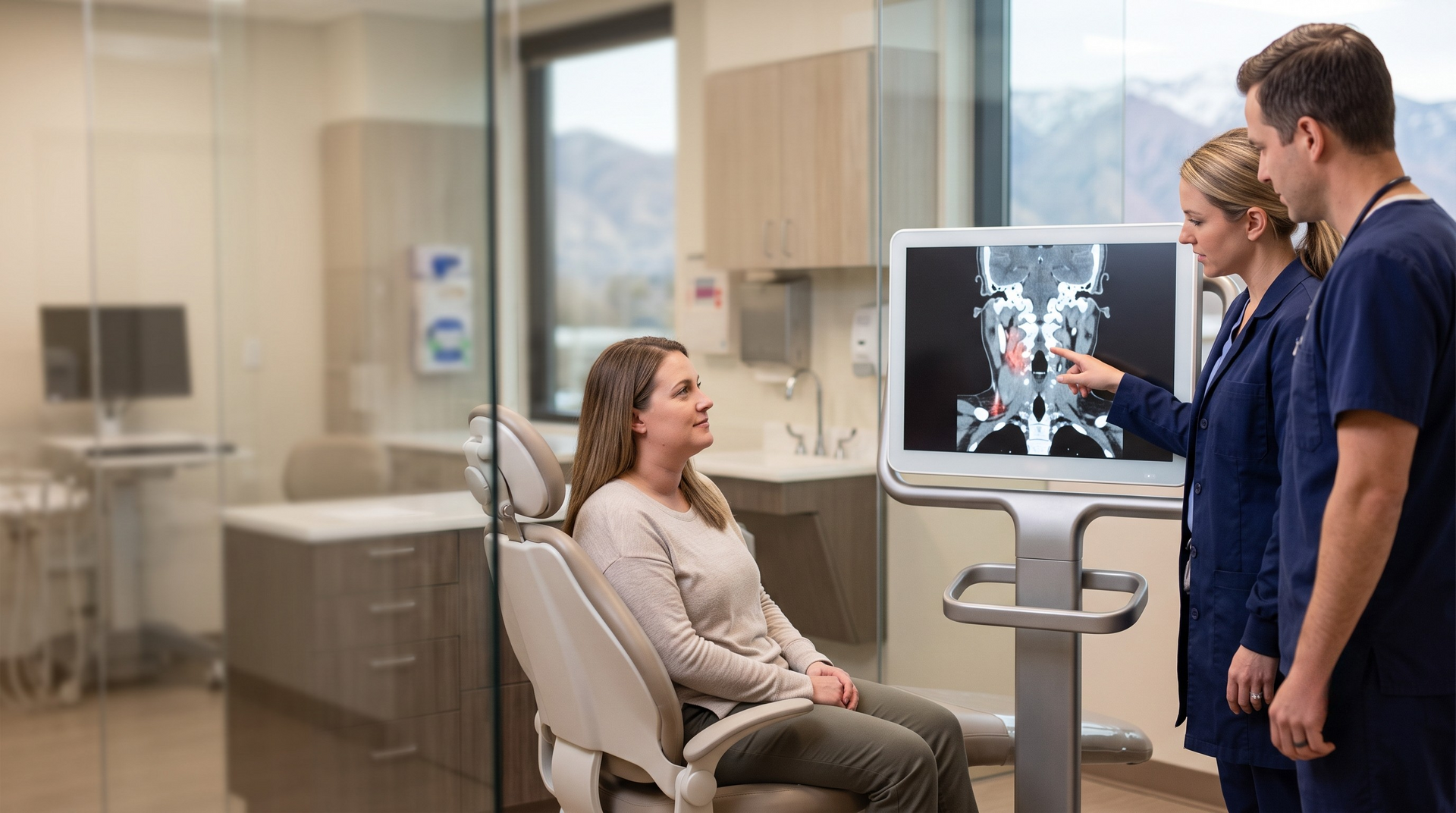
The right procedure depends on what is causing the pain. That is why a dental evaluation and X-ray matter. Once the source is identified, treatment can focus on stopping the problem instead of only masking symptoms.


Common emergency dental procedures include:

  • Root canal treatment: If infection or inflammation has reached the nerve inside the tooth, a root canal can remove the infected tissue, relieve pressure, and help save the tooth.
  • Dental extraction: If a tooth is too damaged or infected to save, removing it can stop the source of infection and pain.
  • Abscess drainage: When appropriate, draining an abscess can reduce pressure and help control infection.
  • Crown or temporary stabilization: A cracked or broken tooth may need protection quickly so the fracture does not worsen.
  • Filling or lost-crown repair: Exposed tooth structure can become painful fast. Restoring coverage can reduce sensitivity and protect the tooth.


These are the kinds of treatments that can change the course of the problem. Pain medication may help you get through a few hours, but dental treatment addresses why the pain is happening.


How Tooth Problems Can Start Affecting the Rest of the Body

Your mouth is connected to the rest of your body through blood vessels, nerves, soft tissues, and the airway. When dental infection spreads beyond the tooth, symptoms can begin to show up outside the mouth.


Warning signs can include:

  • Swelling that moves into the cheek, jaw, neck, or under the tongue
  • Fever, chills, or feeling generally ill
  • Difficulty opening the mouth
  • Trouble swallowing
  • Breathing changes or tightness in the throat
  • Dizziness, confusion, or weakness


These symptoms should be taken seriously. They can suggest that infection or inflammation is no longer limited to one tooth. If breathing, swallowing, or alertness is affected, go to the ER immediately.


When to Call an Emergency Dentist First

If you have severe dental pain but you are breathing normally, swallowing normally, and do not have rapidly spreading swelling or serious whole-body symptoms, an emergency dentist is often the best first call. The goal is to treat the source before it turns into something larger.


Call for same-day emergency dental care if you notice:

  • Throbbing tooth pain that is getting worse
  • Pain that wakes you up or keeps you from sleeping
  • A cracked, broken, or knocked-out tooth
  • A pimple-like bump on the gum
  • Swelling around one tooth or one side of the face
  • A lost filling or crown with sharp pain or sensitivity
  • Pain when biting that feels new or intense


These symptoms do not always mean the worst case scenario, but they do mean the tooth should be checked quickly. The earlier the problem is treated, the more options you may have.


Early Treatment Can Be Less Scary Than Waiting

People often delay care because they are worried about cost, fear, or being told they need a major procedure. That is understandable. But waiting can make the eventual visit more stressful, not less. A tooth that might have needed a filling can progress toward a root canal. A tooth that might have been saved can become an extraction. A localized infection can become swelling that sends you to the ER first.


Early care gives the dental team a chance to explain what is happening, control pain, and choose the most conservative appropriate treatment. It can also help you avoid spending hours in a hospital waiting room only to be told you still need a dentist. If dental pain is escalating, do not wait for it to become unbearable. Same-day emergency dental treatment can help stop the problem before it affects more than your tooth.


What to Do While You Arrange Care

While you are waiting to be seen, keep things simple and safe. Rinse gently with warm salt water, keep your head elevated, avoid chewing on the painful side, and follow the label directions for over-the-counter pain relievers you can normally take. Do not place aspirin directly on the gum or tooth, and do not poke, squeeze, or try to drain a swollen area yourself.

Watch your symptoms closely. If swelling spreads quickly, if swallowing becomes difficult, if breathing feels different, or if fever and confusion appear, go to the emergency room.


Same-Day Help in Salt Lake City

The Emergency Dentist in Murray serves patients across the Salt Lake City metro with same-day emergency dental care, extended evening hours, weekend availability, and Sunday appointments. Walk-ins are welcome, and our team is used to helping people who are scared, tired, and in pain.


Whether the right answer is a root canal, extraction, abscess treatment, repair for a broken tooth, or another procedure, the first step is finding out what is actually causing the pain. Financing as low as 0% interest for 12 months is available through our payment partners, and some plans offer $0 down payments for those who qualify.

If your tooth pain is getting worse, do not wait for it to become an ER-level problem. Call our Salt Lake City area emergency dental team or walk in today so we can help you get relief and protect your health.


You might also like

October 5, 2026
A patient walks into a dental office or an urgent care clinic pointing at the lower jaw. There is no chest pain, no crushing pressure, no radiation down the arm. Just an aching, oppressive jaw or a tooth that will not settle. Most of the time this is exactly what it looks like, an odontogenic problem with a clear dental source. Occasionally it is something far more dangerous: a myocardial infarction announcing itself through the trigeminal map instead of the chest. Recognizing that small subset is one of the highest-stakes pattern-recognition tasks shared by emergency physicians, urgent care clinicians, primary care providers, and dentists.  The data behind cardiac orofacial pain Referred orofacial pain during cardiac ischemia is more common than most clinicians expect. In a 2020 analysis published in the Journal of Oral and Facial Pain and Headache, Kreiner and colleagues reported that roughly 4 in 10 patients experiencing myocardial ischemia describe orofacial pain, typically oppressive or burning in quality. The pain often sits in the lower jaw and throat rather than the upper face, which fits the historical roots of the word angina, meaning to choke. The dangerous minority is smaller but real. In up to 4 to 6 percent of myocardial infarction patients, orofacial pain is the sole presenting complaint, with no chest pain at all. Women report isolated orofacial presentation more often than men. This is the patient who never thinks to call a cardiologist, because nothing about a sore jaw says heart to a frightened person at home. They book a dental appointment instead. Why this matters: patients who present without chest pain are more likely to be misread and undertreated. The foundational National Registry of Myocardial Infarction data (Canto and colleagues, JAMA 2000) found that about a third of confirmed infarctions presented without chest pain, and those patients had markedly higher in-hospital mortality, in part because they received aspirin, reperfusion, and timely intervention less often. Across chest-pain-absent cohorts, one-year mortality runs roughly two to two and a half times higher than in patients who present typically. Kreiner's own conclusion is blunt: a toothache as the only symptom of a heart attack carries a major risk of misdiagnosis and death when the cardiac source is missed in a dental chair. Who is most likely to present atypically Three groups account for most silent or atypical infarctions, and all three are overrepresented among patients whose only complaint is in the jaw or teeth. Women. In the JAMA 2012 registry analysis, 42 percent of women versus 30.7 percent of men presented without chest pain. Younger women carried some of the highest mortality, precisely because the presentation was not taken seriously fast enough. Patients with diabetes. Autonomic neuropathy blunts ischemic pain perception. In the GRACE data, diabetic patients were roughly half as likely to report ischemic chest pain. The warning system is dampened, so the infarction speaks softly, sometimes only through referred jaw pain. Older adults. Each additional year of age lowers the odds of classic chest pain. Patients over 75 are far more likely to present with dyspnea, fatigue, confusion, syncope, or pain referred to the jaw, neck, or back. The pain pattern that should raise the alarm Cardiac-referred orofacial pain has a fingerprint that is almost the mirror image of true dental pain. The single most useful discriminator is what provokes and relieves it. Exertional, not thermal. Cardiac pain comes on with physical effort such as climbing stairs or carrying groceries, and eases with rest. A genuine toothache does not care whether the patient is walking uphill; it responds to hot, cold, biting, and percussion. Often bilateral. Cardiac craniofacial pain is frequently felt on both sides, for example both angles of the mandible. Odontogenic pain is essentially always unilateral and tied to one tooth or quadrant. No dental source on exam. Teeth, alveolar bone, mucosa, and the temporomandibular joint look normal. There is no caries, no fracture, no periapical lesion, and crucially no thermal sensitivity. If cold and percussion testing change nothing, the pain is not coming from the pulp. Company it keeps. Watch for dyspnea, diaphoresis, nausea, light-headedness, or radiation to the arm, neck, or shoulder accompanying the jaw pain. That cluster is the atypical infarction announcing itself. Mechanistically, this is viscero-somatic convergence. Cardiac afferents, including vagal fibers associated with inferior-wall ischemia, share second-order neurons in the upper cervical cord and trigeminal brainstem complex with sensory input from the face and jaw. The brain mislocalizes the threat. The heart is in trouble, but the patient feels a tooth. If a patient describes jaw or tooth pain that flares with exertion, sits on both sides, and produces nothing on a careful dental exam, treat that as a cardiac question first and a dental question second. Our team is available seven days a week to examine the tooth and confirm whether a real dental source exists, so the patient is not left guessing while a serious cause goes unaddressed. The ECG and troponin rule The practical safeguard is simple to state and easy to defend. When orofacial or jaw pain lacks a clear dental source, especially in a patient with cardiac risk factors or an exertional pattern, obtain an electrocardiogram and serial troponin, or refer immediately to a setting that can. Acute coronary syndrome guidance from the American Heart Association and the American College of Cardiology calls for an ECG within 10 minutes of arrival for any presentation suspicious for ischemia, and troponin remains the biomarker that confirms myocardial injury. Missed acute coronary syndrome is far more often a failure to consider the diagnosis than a failure of the test. The threshold to order that ECG should be low in women, older adults, and patients with diabetes. A referral protocol any clinic can use The pathway does not need to be complicated. It needs to be consistent. Examine for an odontogenic source. History, intraoral and extraoral exam, thermal and percussion testing, and imaging where indicated. Identify the offending tooth if one exists. If a clear dental source is found and there are no systemic red flags, proceed with appropriate dental care. If no dental source is found, stop and broaden the differential. Pain that is exertional, bilateral, accompanied by dyspnea or diaphoresis, or occurring in a high-risk patient triggers a same-visit ECG and troponin or an emergency referral, not an empiric procedure. This is where the most preventable harm happens. The literature documents patients who received extractions or root canal treatment for pain that turned out to be cardiac, with the pain unresolved because the tooth was never the problem. One published case describes a 76-year-old woman whose extractions did not relieve her pain, which later worsened with exertion and was finally recognized as cardiac in origin. An irreversible dental procedure performed without an identified odontogenic cause is not a neutral act. It delays the real diagnosis. The mindset: rule out cardiac before drilling The takeaway for every clinician who sees facial and dental pain is a habit of thought more than a protocol. When the tooth does not explain the toothache, the heart earns a seat at the table. No caries, no crack, no periapical lesion, no thermal response, plus an exertional or bilateral pattern in an at-risk patient, means cardiac evaluation comes before any irreversible treatment. The cost of an unnecessary ECG is minutes. The cost of a missed infarction is measured very differently. If you are a clinician trying to clear a tooth quickly so you can safely send a patient onward, or a patient who has been told your jaw pain has no dental cause, we can help confirm whether a real dental source exists. Our practice is open seven days a week, including evenings and weekends, so an aching jaw never has to wait for a weekday appointment to get answered. When the tooth is the problem, we treat it. When it is not, the fastest path to the right care is knowing that early.
July 17, 2026
A patient arrives with a swollen jaw, a few days of throbbing molar pain, and now a fever. The reflex is familiar: antibiotics, analgesia, and a referral note that says "follow up with a dentist." Most of the time that patient is fine. Occasionally that patient is hours away from a lost airway or a mediastinum full of pus. The hard part of odontogenic infection in the emergency department is not treating the obvious abscess. It is deciding which dental infection is quietly behaving like sepsis, and acting before the physiology declares itself.  This is a framework for doing that decisively, built around the same Sepsis-3 lens you already use for pneumonia and pyelonephritis, then layered with the anatomy and source-control realities specific to teeth. The Sepsis-3 lens, applied to the mouth Under the 2016 Sepsis-3 consensus definitions, sepsis is life-threatening organ dysfunction caused by a dysregulated host response to infection. Organ dysfunction is operationalized as an acute rise in the SOFA score of two points or more, a threshold associated with in-hospital mortality above 10 percent. At the bedside, qSOFA flags the patient worth a closer look when two of three are present: respiratory rate 22 per minute or higher, altered mentation, and systolic blood pressure 100 mmHg or lower. Septic shock layers on a vasopressor requirement to maintain a mean arterial pressure of at least 65 mmHg together with a serum lactate above 2 mmol/L after adequate fluid resuscitation, and carries hospital mortality above 40 percent. Here is the honest part. A localized periapical abscess almost never meets Sepsis-3. It is a local infection without systemic organ dysfunction, and treating every swollen face as septic would be both wrong and wasteful. Pure odontogenic sepsis is genuinely uncommon, and death from it is rare. The danger is that the cases which do qualify reach Sepsis-3 through three specific, mechanical or host-dependent pathways rather than through gradual deterioration: Airway obstruction , most classically Ludwig's angina, where bilateral submandibular and sublingual space involvement elevates the floor of the mouth and the tongue. Roughly 90 percent of Ludwig's cases are odontogenic, usually from a mandibular second or third molar. Historical mortality exceeded 50 percent; with modern airway management it is closer to 8 percent. The threat is mechanical, and it can outrun your antibiotics. Descending necrotizing mediastinitis , where infection tracks down the cervical fascial planes into the chest. Mortality is classically reported in the 20 to 40 percent range. This is the patient whose neck and chest pain are not musculoskeletal. Bacteremia in the immunocompromised or poorly controlled diabetic host , where a dysregulated response to a relatively modest dental source produces real organ dysfunction. So qSOFA and lactate still earn their keep here, but a normal qSOFA does not clear a patient with a rising airway threat. The mouth can kill mechanically before it kills systemically. Reading the local exam like a sepsis risk score Treat the oral and neck exam as part of your severity assessment, not a separate dental task. Trismus is a useful proxy for deep-space spread. A practical surgical convention grades it as mild at 20 to 30 mm of interincisal opening, moderate at 10 to 20 mm, and severe below 10 mm; moderate or worse suggests masticator, lateral pharyngeal, or retropharyngeal involvement and should prompt a deliberate airway evaluation. Dysphagia, a muffled or "hot potato" voice, drooling, stridor, and elevation of the floor of the mouth are the findings that move a patient from "dental problem" to "airway emergency." Add the systemic markers you already trust: a high fever, toxic appearance, leukocytosis, elevated CRP, and a lactate above 2 mmol/L as an objective shock signal. When the picture is unclear, a contrast-enhanced CT of the neck is the gold standard for mapping the source and distinguishing drainable abscess from cellulitis. Non-contrast imaging is far less helpful for that distinction.If you take one thing from this piece, make it this: in odontogenic infection the antibiotic is the adjunct and the source control is the cure. A patient sent home on amoxicillin with an untreated necrotic tooth has not been treated. They have been delayed. Building a reliable same-day dental pathway is the single highest-yield thing an ED can do for these patients. Source control: bedside, operating room, and the chair Source control timing matters in the same way it does for any septic focus, and the Surviving Sepsis Campaign emphasizes achieving it as early as feasible. For odontogenic infection, source control has three possible venues. A transoral incision and drainage at the bedside is preferred when the collection is accessible and the airway is secure, because it carries the least morbidity. All but the smallest deep neck abscesses typically warrant operative drainage, and any deep neck abscess with even minimal airway symptoms argues for elective intubation before drainage rather than waiting for the airway to fail. Small abscesses or early phlegmon may settle with aggressive medical management, but if there is no improvement after roughly 48 hours, escalate. The venue your ED cannot provide is the dental chair, and it is often the definitive one. Removing the offending tooth or gaining endodontic access to drain the pulp chamber decompresses the source in a way no antibiotic can. This is exactly where a dependable dental partner changes outcomes: the patient who gets a same-day extraction or pulpectomy is the patient who does not bounce back. Empiric antibiotics that match the microbiology Odontogenic infections are polymicrobial, with early viridans streptococci giving way to anaerobes such as Prevotella and Porphyromonas, and roughly a quarter of isolates penicillin-resistant. For severe or hospitalized disease, ampicillin-sulbactam is a sound first-line empiric choice and has performed well head-to-head against alternatives. Clindamycin retains good bone and anaerobe penetration but carries rising resistance among oral anaerobes and a meaningful C. difficile risk, so it is increasingly hard to defend as monotherapy. For severe presentations escalate to piperacillin-tazobactam or a carbapenem, and add vancomycin or linezolid when MRSA coverage is warranted in the toxic or immunocompromised patient. In penicillin allergy, lean on metronidazole-based regimens, and remember that the old estimate of cephalosporin cross-reactivity is now considered too high. Match the spectrum to severity rather than reaching for the same oral agent for everyone. Disposition: who goes home, who gets admitted A patient can reasonably go home when the collection is localized and drainable, the airway is uninvolved, they are non-toxic and tolerating oral intake, they are immunocompetent, and, crucially, they have a time-bound dental disposition rather than a vague suggestion to find a dentist. Admit for IV antibiotics and involve oral and maxillofacial surgery when there is deep-space involvement, any airway compromise, Ludwig's angina, a necrotizing pattern, immunocompromise, a toxic appearance, or an inability to take fluids by mouth. This matters at a population level too. The United States sees on the order of 1.6 to 2 million non-traumatic dental visits to emergency departments each year, and a large share leave with antibiotics and analgesia but no source control. Around 63 percent of those visits include an antibiotic, which is precisely the pattern that drives repeat visits. The 2019 American Dental Association guideline, endorsed by the American Association of Endodontists, is blunt that antibiotics are not indicated for most pulpal or localized periapical conditions when definitive dental treatment is available. Antibiotic stewardship and good dental disposition are the same goal viewed from two angles. Closing the loop with a same-day dental partner The cleanest fix for the bounce-back cycle is structural: a dental practice that answers the phone, sees urgent referrals the same day, and handles the extraction or endodontic drainage your ED cannot. That relationship turns "discharged with antibiotics and a prayer" into "discharged with a confirmed appointment for definitive source control." Our practice is built for exactly that handoff. We are open seven days a week, including evenings and weekends, and we welcome same-day urgent referrals from emergency departments and urgent care clinicians who need a patient's dental source addressed quickly. If you want a reliable dental partner for your odontogenic infection pathway, we are ready when your patients are.
July 10, 2026
A patient with a bad mandibular molar arrives with neck swelling that looks, at first pass, like cellulitis. The skin is red and tense but not obviously necrotic. The patient is uncomfortable, diabetic, mildly tachycardic, and still talking. It would be easy to order antibiotics, mark the border, and reassess later. Cervical necrotizing fasciitis is the reason that easy path can be lethal. For emergency physicians, infectious disease teams, OMFS, and hospitalists, the critical distinction is this: necrotizing infection often outruns the skin exam. The infection tracks through fascial planes with poor blood supply, so the surface may lag behind the deep tissue process. In odontogenic cases, the source is often a neglected or abscessed mandibular molar feeding polymicrobial infection into the submandibular, parapharyngeal, and cervical fascial spaces. The clock that matters is not the time to the next dose of antibiotics. It is the time to airway control, surgical exploration, and source control.  The odontogenic pattern: small tooth, large field Odontogenic necrotizing fasciitis is rare, but when it appears, it is a high-acuity surgical disease. A systematic review of 164 published odontogenic cases found that every patient required aggressive surgical debridement plus IV antibiotics. Overall mortality was 9.8 percent, but in patients with diabetes it rose to 30.3 percent. That diabetes signal should change triage behavior. A diabetic patient with molar pain, cervical swelling, and systemic toxicity is not just a dental infection with a bigger CBC. The microbiology is usually mixed. Oral anaerobes, streptococci, staphylococci, and gram-negative organisms can all be involved, and cultures often evolve as the disease declares itself. The clinical implication is straightforward: start broad, culture early, narrow later, and do not mistake antibiotics for definitive treatment. Necrotic fascia and necrotic pulp are both poorly penetrated targets. The infected tooth remains a living source of the problem until it is extracted, drained, or otherwise definitively controlled. Why it can look like cellulitis Early necrotizing fasciitis is visually deceptive. The surface can show erythema, heat, swelling, and tenderness, just like uncomplicated cellulitis. The deeper findings are what should make the team stop. Pain out of proportion to the skin findings is the classic clue, but it is not always volunteered clearly, especially in patients who have already taken analgesics or antibiotics. Tenderness beyond the visible erythematous border, rapid progression over hours, systemic findings that feel too big for the skin exam, and a toxic affect are all escalation signs. The signs everyone remembers, dusky skin, bullae, anesthesia, necrosis, and crepitus, are important but often late. In the neck, waiting for the skin to turn dramatic can mean waiting until the airway, mediastinum, or great-vessel neighborhood is already involved. A patient whose neck swelling is becoming firm, spreading, insensate, mottled, or crepitant needs surgical eyes at bedside, not a passive observation plan. LRINEC helps less than people want it to The Laboratory Risk Indicator for Necrotizing Fasciitis score can support suspicion, but it cannot rule this disease out. It was built from lab variables, and necrotizing infection remains a clinical and surgical diagnosis. A low or intermediate score should not calm the room when the history and exam are moving in the wrong direction. Hyponatremia, renal injury, leukocytosis, elevated CRP, lactate, and hyperglycemia can all sharpen concern, but none replaces judgment. Contrast CT is useful when the patient is stable enough to image quickly. In cervical necrotizing fasciitis, CT may show fascial thickening, fluid collections, abscess, edema, and soft-tissue gas. Gas in the cervical fascial planes is a strong warning, but absence of gas does not exclude necrotizing infection. If the patient looks surgically ill, imaging should define the field and airway risk, not delay exploration. If your ED is evaluating a patient with dental-source neck swelling and you need same-day dental source-control coordination after airway and OMFS stabilization, call The Emergency Dentist early. We are open 7 days a week in Salt Lake City and can help close the handoff gap once the surgical team has controlled the deep infection. Treatment is not "IV antibiotics and watch" The treatment pattern is aggressive because the disease is aggressive: resuscitation, airway planning, broad-spectrum IV antibiotics, urgent OMFS or surgical debridement, and repeat exploration when needed. The neck adds an airway problem to the soft-tissue problem. Floor-of-mouth firmness, tongue elevation, dyspnea, dysphagia, drooling, hoarseness, or rapidly expanding submandibular swelling should bring anesthesia, OMFS, and ENT into the room early. If the airway is still manageable, that is the moment to plan it, not proof that it can wait. Empiric antibiotic choices depend on local protocols, allergy history, MRSA risk, and severity, but the principle is broad coverage for oral anaerobes, streptococci, staphylococci, gram-negative organisms, and toxin-mediated disease. Common severe-infection approaches include beta-lactam/beta-lactamase inhibitor therapy or broader gram-negative and anaerobic coverage, plus MRSA coverage when indicated, with clindamycin often used when toxin suppression is a concern. Culture results should guide narrowing, but culture-directed therapy follows source control rather than replacing it. Debridement is the survival intervention Necrotizing fasciitis is named for what surgeons find, not for what the triage note says. Exploration may reveal gray fascia, thin fluid, loss of normal tissue resistance, poor bleeding, necrotic fat, and infected spaces extending beyond the visible skin change. Debridement needs to reach viable tissue, and second-look operations are common because the first operation may not be the last infected edge. Hyperbaric oxygen sometimes enters the discussion. It may be a reasonable adjunct in selected stable patients when available, but the evidence remains mixed and it is not a substitute for debridement. A patient should not be transported away from airway control, resuscitation, and the operating room for an adjunctive therapy while the primary surgical disease is still active. The dental source cannot be an afterthought In odontogenic cervical necrotizing fasciitis, the offending tooth is not incidental history. It is the reservoir. If a mandibular molar with necrotic pulp or periapical infection seeded the neck, the definitive plan should include extraction, endodontic access, or drainage of the dental source in coordination with OMFS. Whenever feasible, source-tooth removal belongs in the same operative episode as fascial debridement, because leaving the dental nidus in place invites ongoing contamination. This is where coordinated referral matters after the acute surgical threat is controlled. The patient may leave the OR with drains, open wounds, antibiotics, and a long recovery path, but the dental pathway still has to be closed. Missed follow-up, delayed extraction, and unclear ownership of the tooth are the quiet ways a catastrophic infection can become a repeat admission. A practical escalation frame Dental source plus systemic toxicity: treat as a deep infection until proven otherwise, especially in diabetes, immunosuppression, older age, renal disease, alcohol use disorder, or malnutrition. Pain out of proportion or rapid progression: call surgery early. Do not wait for bullae or necrosis. Neck firmness, dysphagia, dyspnea, drooling, voice change, or floor-of-mouth elevation: airway planning is the first procedure. Soft-tissue gas or multi-space involvement on CT: move from "possible cellulitis" to urgent operative management. Known molar abscess: document the dental source-control plan before discharge or transfer. Cervical necrotizing fasciitis is uncommon, but the cost of under-calling it is enormous. The safest posture is respectful suspicion: airway first, broad antibiotics early, CT when it helps and does not delay the OR, aggressive OMFS-coordinated debridement, and definitive dental source control. The Emergency Dentist is open 7 days a week in Salt Lake City, including evenings and weekends, and we welcome coordinated handoffs when a dental source needs urgent follow-through after the hospital team stabilizes the life-threatening infection.